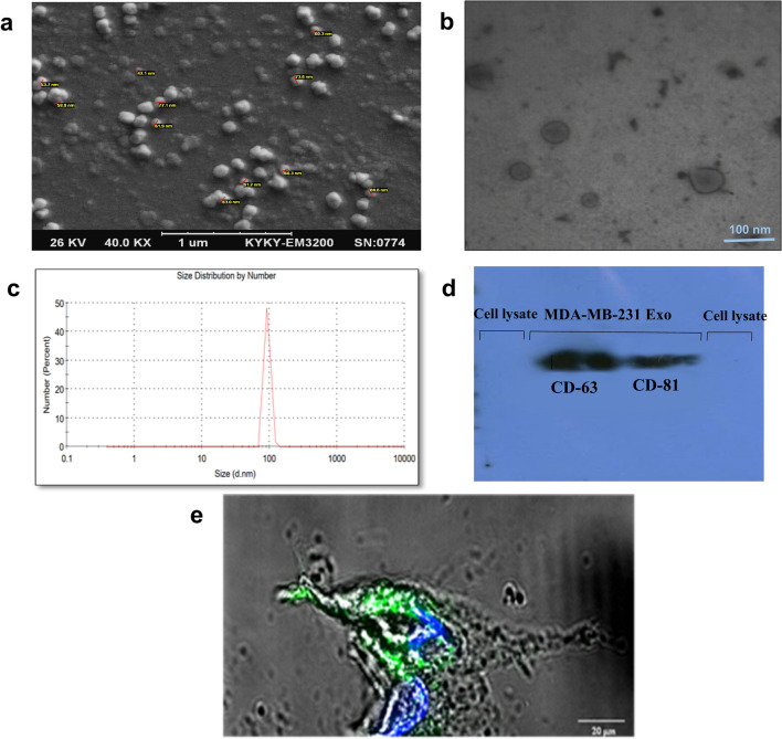
Fig. 1.
Characterization of MDA-MB-231 cell line exosomes and exosome labeling a SEM micrograph of isolated exosomes confirmed the sphere shape and size of exosomes. b TEM micrograph showed the lipid bilayer of exosomes. c DLS analysis based on size distribution by number showed a single peak at ~ 92 nm. d Western blot. Tumor-derived exosomes were positive for CD63 and CD81 as exosomal markers. MCF7 cell lysates were used as a negative control. e Exosomes were labeled with a green fluorescent dye (PKH67), and macrophages were incubated with PKH-labeled exosomes for 24 h. Confocal laser scanning microscope showed the internalization of labeled exosomes in the cytoplasm and around the macrophage nucleus

