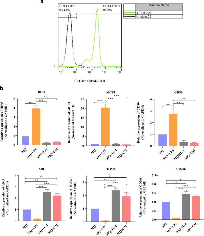
Fig. 2.
Characterization of human peripheral blood monocytes, M1, M2, and tumor-associated macrophages. a The results of flow cytometry analysis confirmed that most of the purified cells are positive for CD14. b After treatment of macrophages by LPS, the results confirmed increased expression of M1 markers (IRF5, MCP1, and CD80) and decreased expression of M2 markers (ARG, TGM2, and CD206) while treatment with IL-4 or conditioned medium of MDA-MB-231 cells increased expression of M2 markers (ARG, TGM2 and, CD206) and decreased expression of M1 markers (IRF5, MCP1, and CD80). *P < 0.05, **P < 0.01

