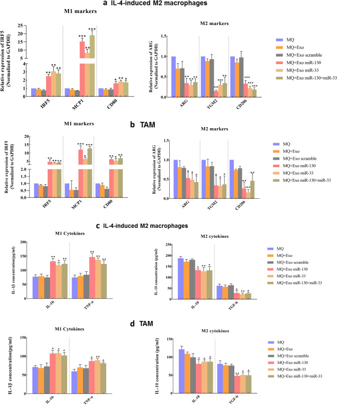
Fig. 4.
The expression of the M1 and M2 specific markers and the production of cytokines. MiRNA-loaded exosomes direct macrophages to a M1 phenotype. IL-4-induced M2 macrophages and TAMs were treated with miRNA-loaded exosomes, and RT-PCR was performed after 48 h. The expression level of M1 markers, including IRF5, MCP1, and CD80 increased while, the expression level of M2 markers, including ARG, TGM2, and CD206, decreased after treatment with miR-130, miR-33, and miR-130 + miR-33-loaded exosomes in both a IL-4-induced M2 macrophage and b TAM. ELISA assay. The cytokine secretion was measured in the conditioned medium of treated macrophages with miRNA-loaded exosomes in c IL-4-induced M2 macrophage and d TAMs. The production level of M1 cytokines (IL-1β and TNF-α) increased while the production level of M2 markers (IL-10 and TGF-β) decreased. *P < 0.05, **P < 0.01, ***P < 0.001

