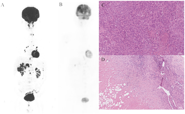
Fig. 2.
A representative case. The patient underwent laparoscopic biopsy in December 2019. PET-CT and H&E staining are shown in 2A and 2C, and pathology revealed a median differentiated gallbladder carcinoma. The patient regularly received 8 mg/day lenvatinib and 200 mg camrelizumab every 3 weeks. Then, in May 2020, PET-CT (2B) showed that the lesions had obviously shrank, and a conventional operation was performed. H&E staining of the surgically resected specimen showed pCR. PET-CT, positron emission tomography computed tomography; H&E, hematoxylin and eosin; pCR, pathological complete response

