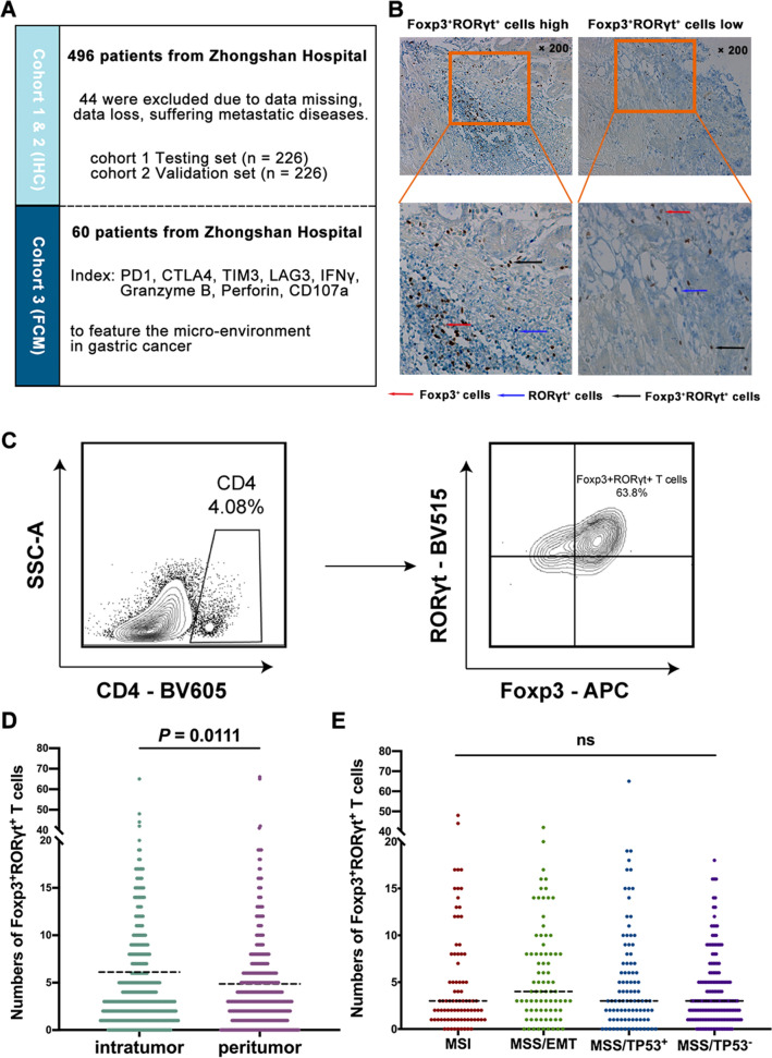
Fig. 1.
Foxp3+RORγt+ T cells are accumulated in gastric cancer and independent of ACRG classification. a The brief summary of the current study. b Immunohistochemistry (IHC) staining of Foxp3+RORγt+ T cells in gastric cancer tissues (intratumor and peritumor). Magnification: × 200. Differenet colors represent corresponding cell subtypes as tagged. c The typical flow cytometry image of Foxp3+RORγt+ T cells was displayed. d IHC score of intratumoral and peritumoral Foxp3+RORγt+ T cells (n = 452). e Association between intratumoral Foxp3+RORγt+ T cells and ACRG classification. Dotted horizontal lines indicate the mean (± SD). NS refers to not significant (one-way ANOVA followed by Tukey’s multiple comparison test)

