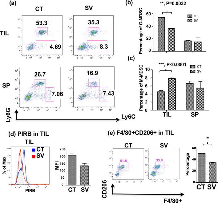
Fig. 4.
SV treatment promotes the acquisition of an M1 phenotype by M-MDSC. a Mice were fed control or SV food starting 10 days prior to tumor inoculation. BALB/c mice were subcutaneously inoculated with 1 × 10⁴ L1 lung cells in 0.1 ml of PBS on day 10 and remained on their corresponding diets. Mice were sacrificed on day 28. Tumor infiltrated leukocytes and splenocytes (SP) were isolated from tumor-bearing mice. G-MDSC (Ly6G+Ly6C+) and M-MDSC (Ly6G−Ly6C+) in TIL and SP were analyzed by flow cytometry and presented as dot plots. b, c Statistical analysis of percentages of G-MDSC (b) and M-MDSC (c) obtained from (a) is shown. **p < 0.01, ***p < 0.001, ANOVA. (d) Phenotypic analysis of PIRB expression in CD11b+ TIL is presented in histogram (left) and as statistical analysis (right). *p < 0.05, ANOVA. (e) The percentage of F4/80+CD206+ M2-type macrophages in TIL is shown in dot plot (left) and the statistical analysis is presented (right). *p < 0.05, ANOVA. Data were presented as the average ± SEM. n = 3 mice per group

