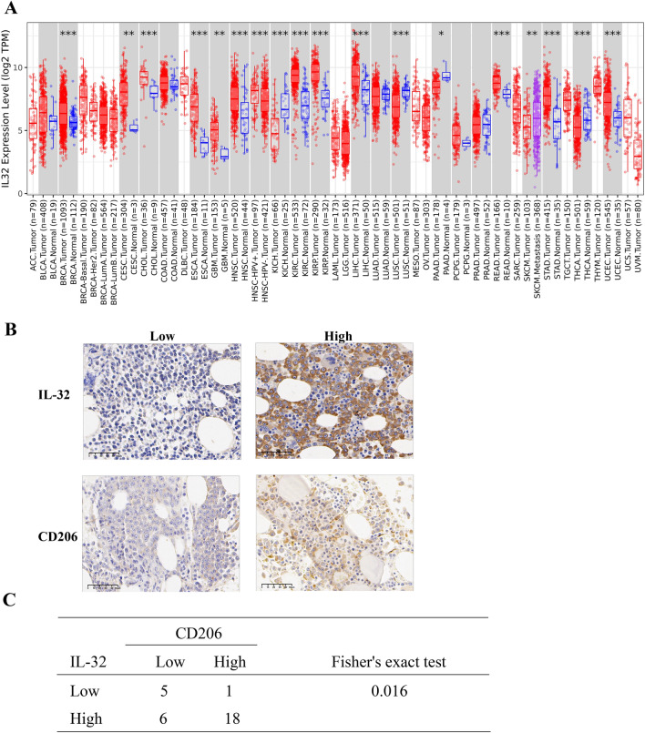
Fig. 1.
IL-32 is highly expressed in relapsed MM patients and correlated with CD206+ M2 MΦ infiltration. a IL-32 mRNA expression was abnormal in various cancer cells as determined by TIMER database analysis. ACC Adrenocortical carcinoma; BLCA Bladder Urothelial Carcinoma; BRCA Breast invasive carcinoma; CESC Cervical squamous cell carcinoma and endocervical adenocarcinoma; CHOL Cholangiocarcinoma; COAD Colon adenocarcinoma; DLBC Lymphoid Neoplasm Diffuse Large B-cell Lymphoma; ESCA Esophageal carcinoma; GBM Glioblastoma multiforme; HNSC Head and Neck squamous cell carcinoma; KICH Kidney Chromophobe; KIRC Kidney renal clear cell carcinoma; KIRP Kidney renal papillary cell carcinoma; LAML Acute Myeloid Leukemia; LGG Brain Lower Grade Glioma; LIHC Liver hepatocellular carcinoma; LUAD Lung adenocarcinoma; LUSC Lung squamous cell carcinoma; MESO Mesothelioma; OV Ovarian serous cystadenocarcinoma; PAAD Pancreatic adenocarcinoma; PCPG Pheochromocytoma and Paraganglioma; PRAD Prostate adenocarcinoma; READ Rectum adenocarcinoma; SARC Sarcoma; SKCM Skin Cutaneous Melanoma; STAD Stomach adenocarcinoma; TGCT Testicular Germ Cell Tumors; THCA Thyroid carcinoma; THYM Thymoma; UCEC Uterine Corpus Endometrial Carcinoma; UCS Uterine Carcinosarcoma; UVM Uveal Melanoma. b Representative immunohistochemistry images of IL-32 and CD206 in BM biopsies from MM patients. Scale bars, 50 μm. c Positive correlation between the expression of IL-32 and CD206 based on Fisher’s exact test. Samples from 30 MM patients were used

