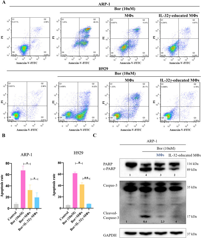
Fig. 2.
IL-32γ enhances the protective effect of MΦs on MM cells. a MΦs were stimulated with IL-32γ for 24 h and then cocultured with MM cells (ARP-1 or H929 cells), and Bor was added for 24 h to induce apoptosis. A representative flow cytometry analysis shows the apoptosis of MM cells. b Summarized results from at least three independent experiments. Values are presented as means ± SEM. c Western blotting was used to detect apoptotic proteins (c-PARP and c-Caspase3) in ARP-1 cells. The quantified density is shown below the bands. The data are representative of at least three independent experiments with similar results. *p < 0.05, **p < 0.01

