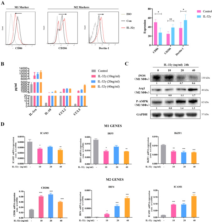
Fig. 3.
IL-32γ induces the polarization of M2 MΦs. a MΦs were stimulated with IL-32γ (20 ng/mL) for 24 h, and flow cytometry was used to detect the expression of CD86, CD206 and Dectin-1 on the surface of the MΦs. The summarized results are from at least three independent experiments. Values are presented as means ± SEM. b MΦs (biological samples from two donors) were stimulated with IL-32γ (10, 20 or 40 ng/mL) for 24 h, and a cytokine chip was adopted to detect the expression of IL-1a, IL-10, CCL2 and CCL5 in the cell culture supernatant. c Western blotting was used to detect the expression of iNOS, Arg1 and p-AMPK. The quantified density is shown below the bands. The data are representative of at least three independent experiments with similar results. d qRT-PCR was adopted to evaluate the expression of M1 and M2 MΦ-related molecules. Data are presented as means ± SEM of at least three independent experiments. *p < 0.05, **p < 0.01, ***p < 0.001

