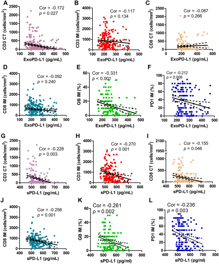
Fig. 3.
Preoperative exoPD-L1 and sPD-L1 showed strong degree of correlation with activate immune status in colorectal liver metastasis sites. Preoperative exoPD-L1 showed high correlation with A CD3 + T cell infiltration at the tumor center (CT) (Spearman’s correlation at P = 0.027, r = − 0.170), E GB expression at the invasive margin (IM) (P < 0.001, r = − 0.331), and F PD1 expression at CT (P = 0.008, r = 0.212), but no correlation with B CD3 + T cell infiltration in invasive at CT (P = 0.134, r = 0.117) and C CD8 + T cell infiltration at CT (P = 0.266, r = -0.087) and D CD8 + T cell infiltration at IM (P = 0.240, r = − 0.092); preoperative sPD-L1 showed high degree of correlation with G CD3 + T cell infiltration at the tumor center (CT) (Spearman’s correlation at P = 0.003, r = − 0.228), H CD3 + T cell infiltration at the invasive margin (IM) (P < 0.001, r = − 0.270), I CD8 + T cell infiltration at CT (P = 0.046, r = − 0.155), J CD8 + T cell infiltration at IM (P = 0.001, r = − 0.256), and K GB expression at IM (P = 0.002, r = − 0.261), and (L) PD1 expression at IM (P = 0.003, r = − 0.236)

