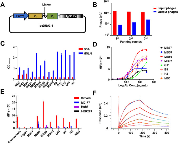
Fig. 1.
Selection and expression of anti-MSLN scFv antibodies by phage display. A Scheme of phagemid vector pcDNA3.4 used for scFv display. B Enrichment of phage-displayed scFvs after each round of panning, and the output phages will be used for monoclonal screening. C Screening of positive clones bind to hMSLN by monoclonal phage ELISA. Each phage-displayed scFv was tested against hMSLN or BSA. D The antibody (ug/ml) with serial twofold dilutions was mixed and incubated with tumor cells expressing MSLN. Characterization of six most potent binders by noncompetitive ELISA. SPR analysis of MSLN over a range of concentrations. E Detection of MSLN antibody specificity by flow cytometry. Ovcar3, a MSLN-positive cell line. MC-F7, Huh7 and HEK293 are MSLN-negative cell lines. F BLI sensor grams of G11cloned binding to biotin-labeled MSLN immobilized on SA chip

