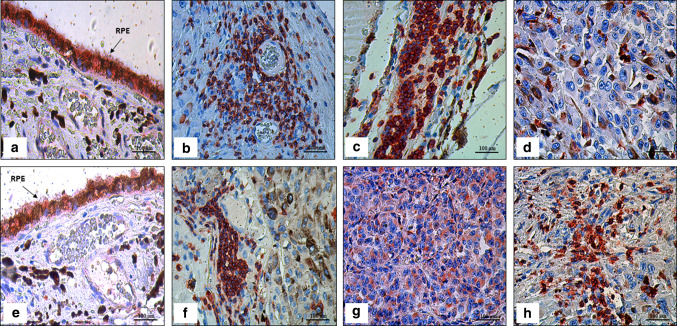
Fig. 1.
Immunoexpression in uveal melanoma (UM) tissue samples with and without tumor infiltrating lymphocytes (TILs) using AEC (3-amino-9-ethylcarbazole) red chromogen, and then counterstained with hematoxylin. a Immunoexpression of PD-1 in retinal pigment epithelium (RPE; arrow) as an internal control (× 400); (b) Immunoexpression of PD-1 in TILs near the intratumoral blood vessels (× 400); (c) Immunoexpression of PD-1 in TILs (× 400); (d) Weak immunoexpression of PD-1 in mixed cell type (× 400); (e) Immunoexpression of PD-L1 in retinal pigment epithelium as an internal control (× 400); (f) Immunoexpression of PD-L1 in TILs (× 400); (g) Cytoplasmic immunoexpression of PD-L1 in epithelioid UM (× 400); (h) Strong immunoexpression of PD-L1 in mixed cell type (× 400)

