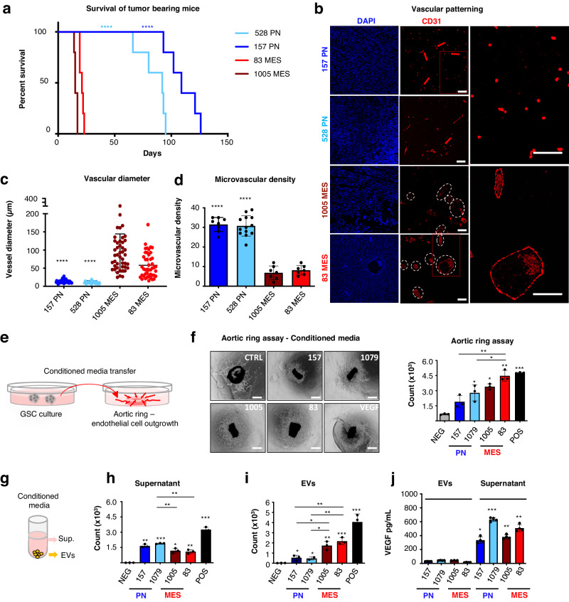
Fig. 1. Differential vascular patterns in GSC-driven tumours and vascular activities of soluble and vesicular components of glioma stem cell secretome.
a Kaplan-Meier survival curves of mice bearing PN GSC- and MES GSC-derived tumours. (n = 5 independent experiments. Two-tailed paired t test. P = 0.0000965 and 0.000089) b Representative images of immunofluorescence for CD31 reveal phenotypic vascular differences between tumours driven by PN or MES GSCs. (n = 5 independent experiments). c Quantification of vessel size distribution through tracing CD31 positive endothelial cells. Blood vessels in MES tumours present enlarged lumens, up to 90 μm, compared to a mean vessel diameter of 13 μm in the PN tumours (n = 5 mice/group. Two-tailed paired t test P = 5.48−16 and 1.18−12). d Quantification of microvascular density using CD31 staining (n = 5/group. Two-tailed paired t test. P = 5.25−09 and 2.68−09). Microvascular density was expressed as vessel density per high power field (hpf). Scale bars are 50 µm e Schematic diagram illustrating mouse aortic ring endothelial outgrowth assay using conditioned media derived from different glioma stem cells. f Endothelial responses induced by the GSC conditioned media containing soluble fraction of the secretome and EVs. RhVEGF was used as positive control [25 ng/mL]. Cells were imaged with optical microscope (left) to assess the number of endothelial cells growing out of the ring using FIJI software (right) (n = 6 independent experiments. Two-tailed paired t test. MES P = 7.55−05 and 2.65−04). g Schematic diagram illustrating secretome fractions preparation using centrifugation methods. h Endothelial responses induced by the GSC secretome. Mouse aortic rings were seeded under domes of BME and cultured in growth factors-enriched media. After rings began to form endothelial outgrowth (often referred to as ‘sprouts’) they were treated with supernatant fractions (PN P = 0.00037 and 3.31609−06. MES P = 0.0010 and 0.00073) or with (i) 30 μg/mL of EVs obtained from either PN GSC (157;1079), or MES GSC (83; 1005). RhVEGF was used as positive control [25 ng/mL]. The number of endothelial cell outgrowths from the ring was assessed using FIJI software. (n = 6 wells/3 independent experiments. Two-tailed paired t test. PN P = 0.0229 and 0.0023. MES P = 0.0021 and 0.00037). j VEGF content distribution in EVs and in supernatant fractions of GSC conditioned media. ELISA assay quantification showed that the growth factor is virtually absent in EVs, and preferentially released in soluble form into the culture media supernatant (n = 4 independent experiments. Two-tailed paired t test. PN P = 1.21−05 and 1.30−08. MES P = 8.08−06 and 9.83−07). Results are shown as mean ± SD; *p < 0.05, **p < 0.01, ***p < 0.001 treated group versus control group ****P < 0.0001; Detailed data are provided as a Source Data file.

