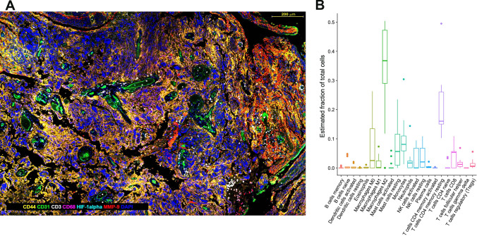
Fig. 2.
a Multiplex IHC using opal immunohistochemistry. GBM (WHO grade IV astrocytoma) tissue was stained with antibodies against CD44 labels tumor cells; CD31 labels endothelial cells (blood vessels); CD68, labels macrophages; HIF-1alpha, labels hypoxic cells; MMP-9 labels matrix metalloproteinase-9-producing cells; DAPI labels cell nuclei. Software-aided image analysis allows all single- and multiplex-labeled cells to be identified and counted, and the relative distances between cell types and histopathological hallmarks to be measured. Scale bar is 200 μm. b Immune cell composition in GBM inferred by computational deconvolution using CIBERSORT. GBM RNAseq data from the 168 GBM patient samples (The Cancer Genome Atlas (TCGA) [35]), was analyzed using the CIBERSORT algorithm to estimate the number of immune cells in GBM. Each data point represents an individual patient tumor sample

