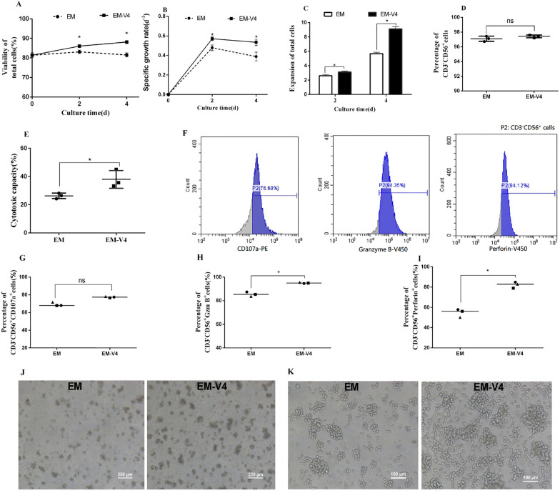
Fig. 2.
Ex vivo expansion characteristics of NK-92 cells. A Cell viability. B Specific growth rate of NK-92 cells. C Expansion fold of the cells. D Percentage of CD3−CD56+ cells. E Cytotoxicity of expanded NK-92 cells on day 4 at an E:T ratio of 5:1. F Representative flow cytometric analysis of CD107a+, granzyme B+ and perforin+ cells which grew in EM-V4; G Percentages of CD107a+ cells gated on CD3−CD56+ cells; H percentages of granzyme B+ cells gated on CD3−CD56+ cells; I percentages of perforin+ cells gated on CD3−CD56+ cells. J Light microscopic image of NK-92 cell morphology at 40 times magnification on day 4. K Light microscopic image of NK-92 cell morphology at 100 times magnification on day 4. (*P < 0.05, n = 3)

