FIGURE 4.

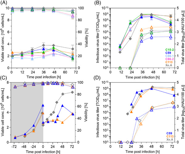
Screening of MDCK cell clones for A(H1N1) production in an ambr15 system and in 1 L STRs. Cultures with 2 × 10⁶ cells/mL in trypsin‐containing medium were infected with A/PR8 at MOI 10−3 under standard conditions (triangles: clones in MDXK medium, diamond‐shape, gray: Xe.A in 4Cell medium). Cells were (A) directly seeded with target VCC in the ambr15 vessels, or (C) first grown in a 1 L STR system and subsequently diluted at time of infection. VCC (full symbols, solid lines) and cell viability (dashed lines, empty symbols); infectious (full symbols, solid lines) and total virus titer (dashed lines, empty symbols) determined by TCID50 and HA assay, respectively. (A, B) Mean and standard deviation of three culture vessels; 1 L STR (C, D): single runs. Gray area in (C) represents cell growth phase not shown in the other graphs.
