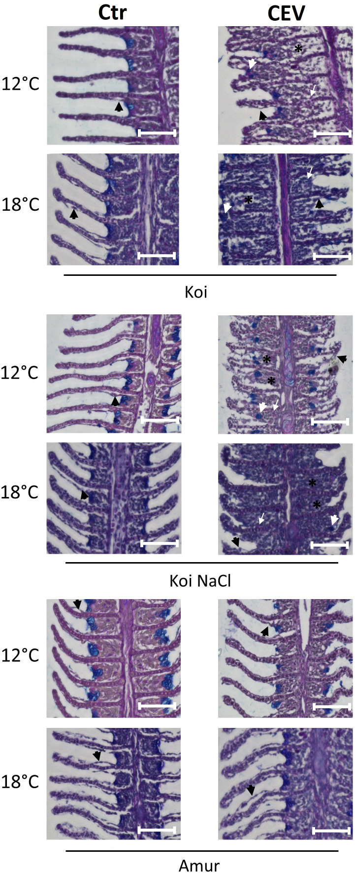
Figure 2.
Histology of the gills during CEV infection (6 days post exposure). Black arrowheads indicate examples of lifting of the epithelial cells on the secondary lamella. White arrowheads indicate examples of translocated goblet cells. Black (*) symbol indicates examples of occluded interlamellar space. White arrows indicate examples of macrophage infiltration. Bar 100 µm. AB-PAS staining.

