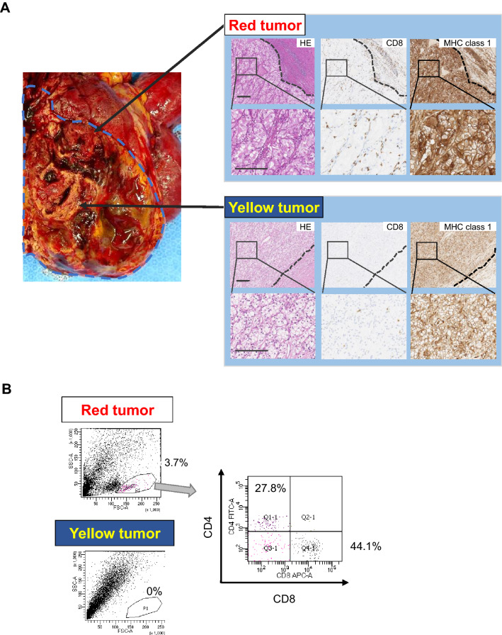
Fig. 1.
Macroscopic, pathological, and immunological findings of a clear cell RCC case. a Macroscopic and microscopic images of the tumor. Macroscopically, the tumor showed heterogeneity with a reddish lesion (red tumor) and yellowish lesion (yellow tumor). An RCC cell line, 1226 RCC cells, was established from the red tumor. The yellow tumor showed clear cell RCC with Fuhrman nuclear grade 1, and the red tumor showed Fuhrman nuclear grade 2. CD8+ T cell infiltration and HLA class 1 expression were examined by immunohistochemical staining. Dotted lines indicate the epithelial-subepithelial margin. HE: hematoxylin and eosin staining. Original magnification × 250 (upper image) and × 500 (lower image); bar = 200 μm. b TIL analysis by flow cytometry. Flow cytometry analysis of TILs in the red and yellow tumors. TILs from the red tumor contained 27.8% CD4+ T cells and 44.1% CD8+ T cells in 3.7% of putative lymphocytes gated by forward scatter and side scatter using tumor single cell suspensions. No TILs were detected from the yellow tumor

