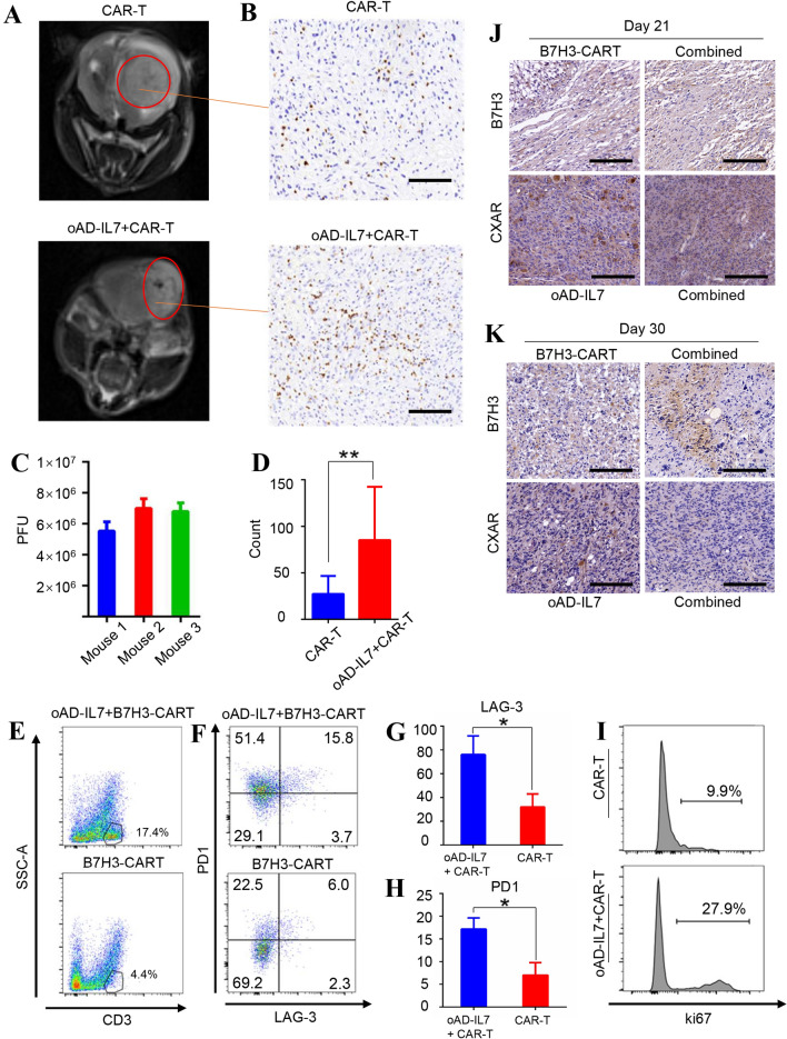
Fig. 5.
oAD-IL7 prolonged the survival of tumor-infiltrating CAR-T cells in xenograft models. In a parallel experiment, the tumor-bearing mice underwent a delayed treatment since the 21st day after tumor inoculation. a The tumors of the mice treated with single B7H3-CAR-T therapy or combined therapy were imaged by MRI scanning at day 28. b The tumors were removed from the treated mice at day 30, and the immune infiltrate was evaluated by immunohistochemical staining according to the distribution of CD3+ cells. Scale bar, 200 μm. c Titration of oAD-IL7 in glioblastoma tissue by plaque assay. d The ratio of CD3 positive cells in tumor tissues removed from mice treated by combined therapy or single B7H3-CAR-T therapy according to the count of average CD3 + cells in each single field. e Representative flow cytometry analysis of the T cells in the tumor removed from mice underwent different therapies. f The expression of PD1 and LAG-3 of the CD3 positive cells in two groups. g The level of LAG-3 expression on the tumor-infiltrating T cells of two groups. h The level of PD1 expression on the tumor-infiltrating T cells of two groups. i Ki67 expression of the CD3 positive cells in two groups. j Expression of the B7H3 and CXAR of the tumors removed from tumor-bearing mice on day 21. Scale bar, 50 μm. k Expression of the B7H3 and CXAR of the tumors removed from tumor-bearing mice on day 30. Scale bar, 50 μm

