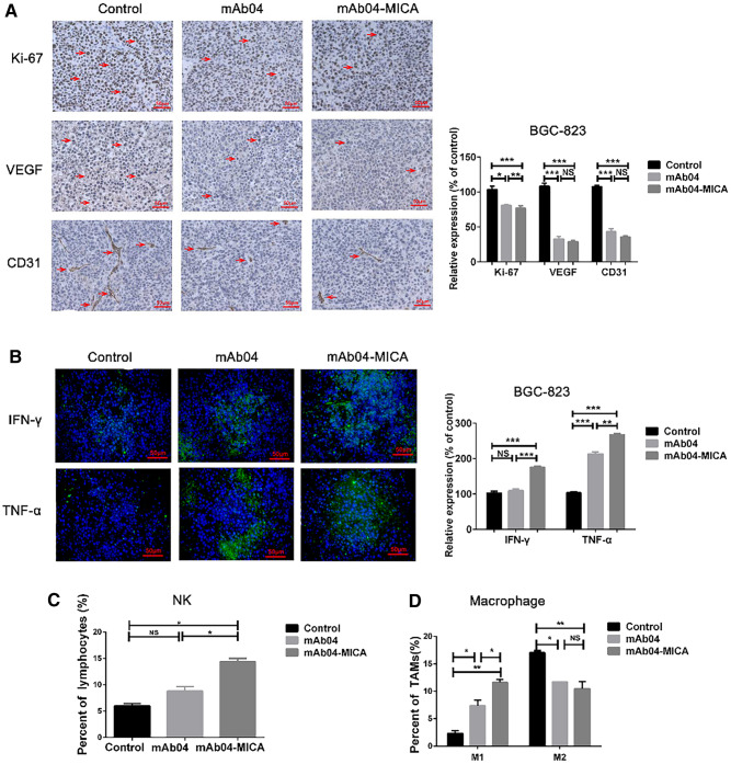
Fig. 4.
mAb04-MICA reduced tumor cell proliferation and angiogenesis, and activated the innate immune system in tumor tissue. A, IHC staining of Ki-67, VEGF, and CD31 on paraffin sections in BGC-823 xenografted tumors. Positive cells were identified with antibodies in brown staining. Quantitative analysis is shown on the right. Ki-67 was counted with Image-Pro-Plus. Relative expression of Ki-67 (%) = (Experiment/Control) × 100%, and the formula is appropriate for calculating the relative expression of VEGF and CD31. Photomicrographs showed representative pictures from three independent tumor samples. Magnification, × 400. B, immunofluorescence staining to evaluate the levels of IFN-γ and TNF-α (green fluorescence) released in different groups. Magnification, × 400. Quantitative analysis is shown on the right. C, representative histogram (obtained from flow cytometry results) of frequency of CD3−CD49b+ NK cells in tumor tissue. D, representative histogram (obtained from flow cytometry results) of frequency of F4/80+CD16/32+ M1 macrophage and F4/80+CD206.+ M2 macrophage in tumor tissue. After administration for 10–12 d, mice were sacrificed, and the immune cells obtained from the tumor tissue were analyzed by flow cytometry. Data were presented as the mean ± SD, n = 3, **p < 0.01, ***p < 0.001

