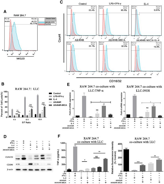
Fig. 5.
AK404R-MICA induced the polarization of macrophages to M1 type in vitro. A, expression of NKG2D on RAW 264.7 detected by flow cytometry. RAW 264.7 was co-cultured with LLC for 24 h before detection. RAW 264.7 cells were co-cultured with LLC for 24 h with 5 ng/mL IFN-γ + 100 ng/mL LPS,10 ng/mL IL-4, 1000 nM AK404R or AK404R-MICA for another 48 h, respectively. Then culture supernatant and the cells were harvested to analysis: B, cytotoxicity assay to assess the RAW 264.7 cell-mediated killing of LLC cells by LDH release assay. C, flow cytometry detected the proportion of CD16/32.+ M1 type macrophages under different conditions. D, western blot analysis for the CD16/32 and CD206 for M1 and M2 type macrophages respectively. E, qPCR analysis for TNF-α and iNOS released by M1 macrophages. F, left, the release of TNF-α detected by ELISA. Right, the release of NO detected by DAF-FM DA. Data were presented as the mean ± SD, n = 3, **p < 0.01, ***p < 0.001

