Fig. 7.
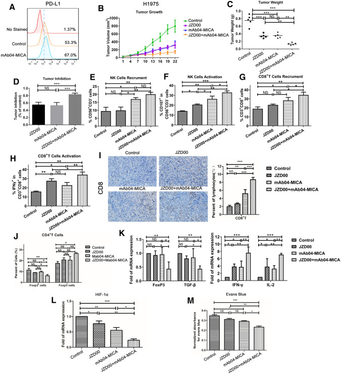
Anti–PD-L1 enhances the anti-tumoral activity of Mab04-MICA in NSCLC model through stimulating infiltration and activation of NKs and CTLs in responding tumors. A After mAb04-MICA treatment, the expression of PD-L1 in tumor tissues was detected by flow cytometry. B Subcutaneous tumor growth curve of H1975 human non-small cell lung cancer mice, C tumor weight, D tumor inhibition rate. (N = 5). Data were given as the mean ± SD (n = 5). *p < 0.05, **p < 0.01, ***p < 0.001. E–J, Recruitment and activation of NKs and CTL in tumor tissue. A NOD-SCID mouse xenografted tumor model of human non-small cell lung cancer was established. After 4–5 doses, flow cytometry was used to detect the infiltration rate of CD56+NK cells(E), number of activated CD107a + CD56 + NK cells(F), CD8 + T cell infiltration level(G), activated IFN-γ + CD8 + T cells(H) in tumor tissues. I. Immunohistochemical detection of infiltrating CD8 + T cells in tumor tissues and statistical analysis. J, CD4 + Foxp3 + Treg and CD4 + Foxp3-Treg. K, the release of Foxp3, TGF-β, IFN-γ, IL-2, HIF-1α, and other cytokines was detected by qPCR. M, Evans Blue detects the degree of vascular leakage

