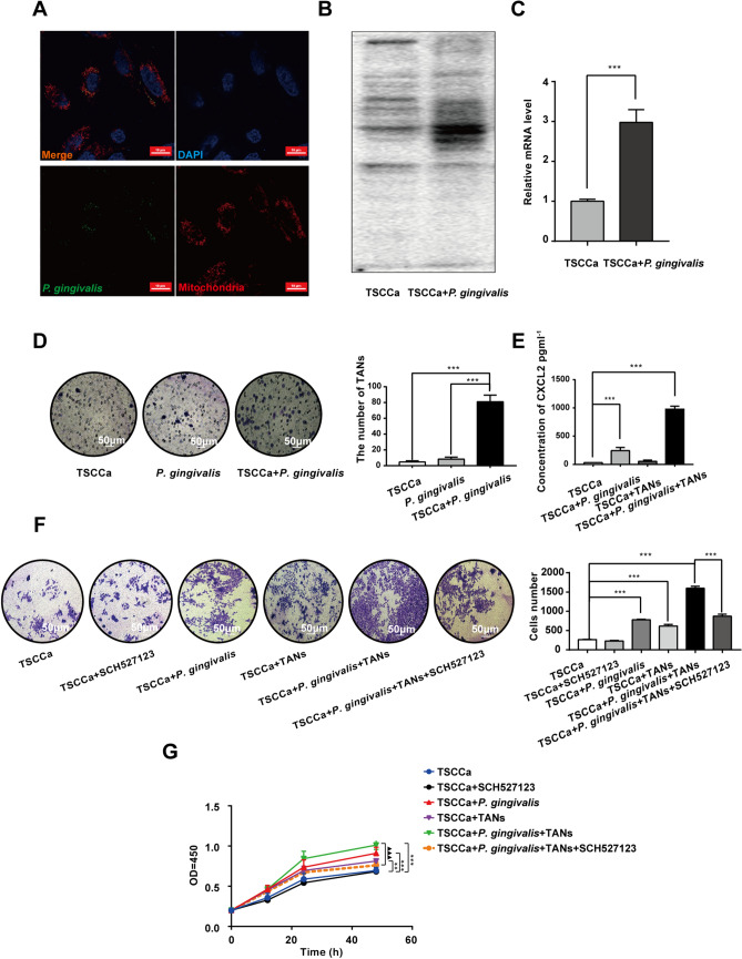
Fig. 4.
The verification of cell coculture models and experiments assessing cell biological behaviour. a P. gingivalis was marked by the fluorochrome of SYTO9, the nucleus was marked by DAPI, and the mitochondria were marked by MitoTracker red. b The difference in the expression of P. gingivalis protein between the coculture cell model and the TSCCa cell model was detected by Western blotting. c The difference in the expression of P. gingivalis mRNA between the coculture cell model and the TSCCa cell model was detected by qRT-PCR. d A neutrophil chemotaxis experiment was used to detect the chemotactic ability of TANs in the supernatants of TSCCa, P. gingivalis, and TSCCa + P. gingivalis culture systems. e The content of CXCL2 in the supernatants of TSCCa, TSCCa + P. gingivalis, TSCCa + TANs and TSCCa + P. gingivalis + TANs coculture systems. f, g Invasion and CCK-8 assays were used to detect the differences in cell invasion and proliferation in the TSCCa, TSCCa + SCH527123, TSCCa + P. gingivalis, TSCCa + TANs, TSCCa + P. gingivalis + TANs and TSCCa + P. gingivalis + TANs + SCH527123 groups (**p < 0.01, ***p < 0.001 and ▲▲▲p < 0.001; one‐way analysis of variance, Student's t-test)

