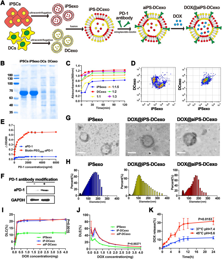
Fig. 1.
Construction and characterization of nanosystems DOX@aiPS-DCexo. A Scheme of the preparation of DOX@aiPS-DCexo. B SDS-PAGE protein analysis of the whole protein expression in iPSCs, DCs, iPSCs exosomes and DCs exosomes. C Determination of optimal fusion ratio of two exosomes. D Flow cytometry analyzer of the fusion of the two nanoparticles. E OD values at 450 nm in different concentrations antigen coated plates. F WB analysis of vector-modified PD-1 antibody. G Transmission electron microscopy (TEM) observation of the surface morphology of DOX@aiPS-DCexo. Scale bars are 100 nm. H DLS analysis of particle size distribution of nanosystem DOX@aiPS-DCexo. Encapsulation rate (I), drug loading capacity (J) and drug release (K) of the nanosystem DOX@aiPS-DCexo. All data were expressed as mean ± SD. Statistical significance was calculated by one-way ANOVA with Tukey’s post hoc test

