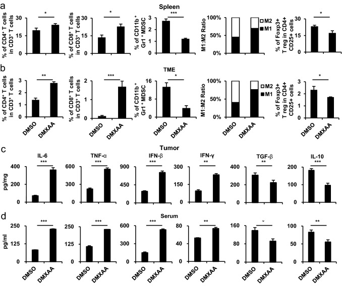
Fig. 2.
Characterization after the co-administration of tumor-specific antigen and STING agonist in cisplatin-treated mouse model. In the in vivo experiments, the groups were as follows: cisplatin treatment with E7 long peptide vaccination (DMSO) and cisplatin treatment with E7 long peptide and DMXAA vaccination (DMXAA). a Tumor mass was measured until the mice died or the tumor diameter reached > 2 cm (n = 5). b Mouse survival was observed for 60 days (n = 5). c One week after the last vaccination, the tumor tissues and spleens of TC-1 tumor-bearing mice were harvested and re-stimulated with E7 short peptide and then analyzed for IFN-γ+ CD8+ T cells by flow cytometry (n = 5). d, e Tumor tissues and spleens of the mice were harvested on day 22. Bar graphs depict the presence of CD4+ T cells, CD8+ T cells, MDSCs, and Treg cells and the M1 and M2 distribution percentages of CD11b+ F4/80+ macrophages, as evaluated by flow cytometry analysis (n = 5). f, g One week after the last vaccination, the tumor tissues and serum from the same mice in c were harvested. Bar graphs represent the levels of cytokines in the tumor tissue and serum of mice as measured by ELISA (n = 5). IBM SPSS Statistics Base 22.0 was used for statistical analysis. *P < 0.05, **P < 0.01, ***P < 0.001

