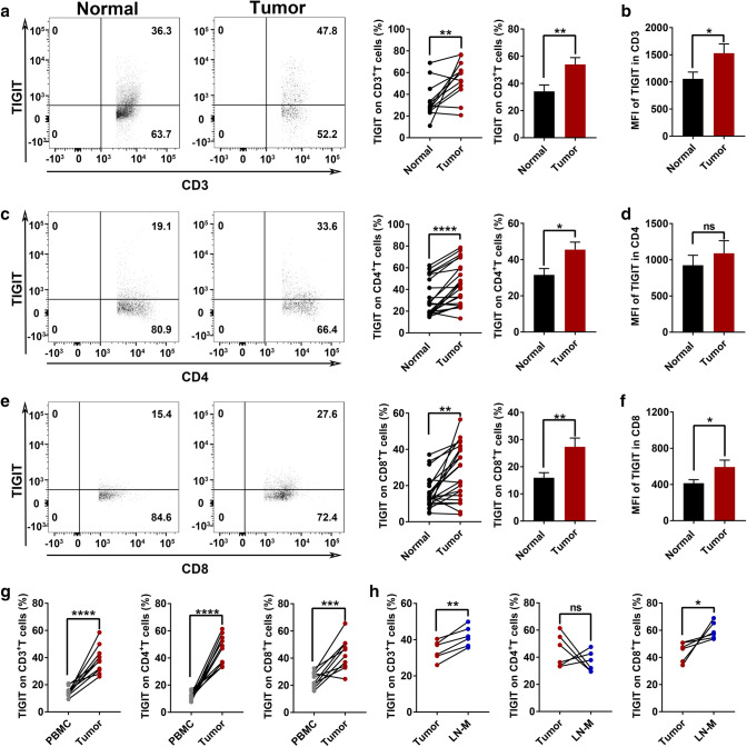
Fig. 1.
TIGIT is upregulated in colorectal cancer TILs and metastases, especially in CD8+T cells a. Different expressions of TIGIT expression in CD3+T cells between normal tissue and tumor tissues. n = 12. b. The mean fluorescence intensity of TIGIT in CD3+T cells. n = 12. c. Different expressions of TIGIT expression in CD4+T cells between normal tissue and tumor tissues. n = 22. d. The mean fluorescence intensity of TIGIT in CD4+T cells. n = 22. e. Different expressions of TIGIT expression in CD8+ cells between normal tissues and tumor tissues. n = 22. f. The mean fluorescence intensity of TIGIT in CD8+T cells. n = 22. g. Different expressions of TIGIT expression in CD3, CD4, and CD8+T cells between PBMC and tumors. n = 10. h. Different expressions of TIGIT expression in CD3, CD4, and CD8+T cells between tumors and lymph node metastases. n = 6. (PBMC, peripheral blood mononuclear cell; LN-M, lymph node metastases). Error bars represent mean ± SEM. Two-tailed Student’s t test and paired t-test were performed for statistical analysis; ns: not significant; *:p < 0.05; **:p < 0.01; ***:p < 0.001; ****: P < 0.0001

