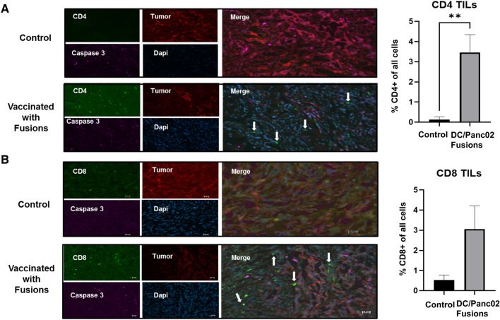
Fig. 6.
Cryosections indicating increased tumor infiltrating lymphocytes (TILs) in DC/Panc02 fusions treated mice compared to control mice. Subcutaneous tumors were removed from a vaccinated and control animal and tissue was fixed in 4% PFA. Cryosections were prepared and stained for CD4 and CD8 positive cells (green), caspase 3 (pink), and DAPI (blue). Tumor cells were identified by staining for M-cherry (red). Cryosection of tumor removed from control mouse indicating a large percentage of tumor cells and relative absence of tumor infiltrating CD4 A and CD8 B T cells. In contrast, vaccinated animals demonstrated relative paucity of tumor with presence of infiltrating CD4 A and CD8 B T cells. Bar graphs depict the percentage of CD4 TILs as quantified by the average value from enumeration of cells in five independent fields

