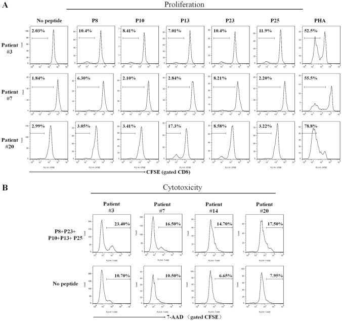
Fig. 2.
The 5 validated epitopes induced CD8+ T cell proliferation and cytolysis of patients’ PBMCs in vitro. The PBMCs from three HLA-A0201+/GPC3+ HCC patients were prelabeled with CFSE and co-cultured with each validated epitope peptide for 7 days followed by flow cytometry to detect the proliferation frequency of CD8+ T cells. a The proliferation profiles of CD8+ T cells in response to each epitope peptide, PHA (positive control) or no peptide (negative control) for each patient’s PBMCs. In parallel, the PBMCs from four HLA-A0201+/GPC3+ HCC patients were co-cultured with the cocktail of 5 validated epitope peptides for 7 days, then cells were harvested and co-cultured as effector cells with K562 cells and CFSE-prelabeled HepG2 cells for 4 h. After 7-AAD staining, cytotoxicity was measured by flow cytometry. b Cytolysis profiles and the frequencies of 7-AAD+ cells in the CFSE+ cell populations at an E: T ratio of 30:1

