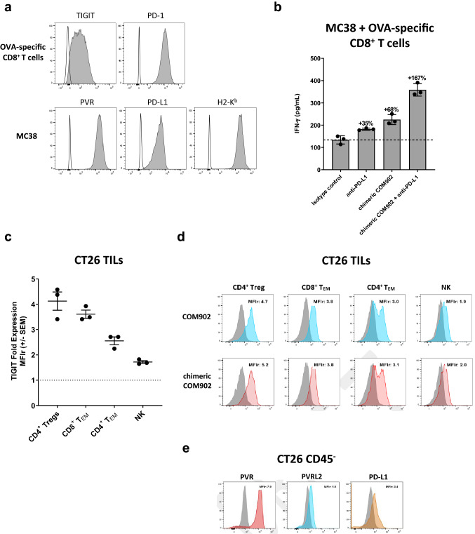
Fig. 7.
Chimeric COM902 enhances in-vitro anti-tumor activity of mouse T cells. a The expression of TIGIT and PD-1 on OT-1 splenocytes activated for 3 days with OVA(257–264) peptide and rhIL-2, as well as of H-2Kb, PVR, and PD-L1 on MC38 target cells was assessed. Gray histograms represent the target staining and white histograms represent staining with the matched isotype control antibodies. Plots shown are from a representative experiment (n = 2). b Mouse OVA-specific CD8+ T cells were co-cultured with OVA-peptide-pulsed MC38 cells in the presence of the chimeric COM902 as a monotherapy or in combination with PD-L1 blockade. The secretion of IFN-γ relative to isotype control is shown for each condition. Mean ± SD are shown by the hash marks. c TIGIT expression on infiltrating lymphocytes isolated from s.c. CT26 tumors is shown as fold expression relative to the isotype control antibody (MFIr) for each cell subset. Each dot represents an individual mouse (n = 3) and mean ± SEM. d Representative TIGIT expression on indicated cell populations from CT26 tumors. Blue, red, and gray histograms represent staining with COM902, chimeric COM902, and the mIgG1 isotype control, respectively. e PVR, PVRL2, and PD-L1 expression on CD45- cells isolated from CT26 tumors. Gray histogram represents mIgG1 isotype control. Representative MFIr values are reported to the right of each histogram

