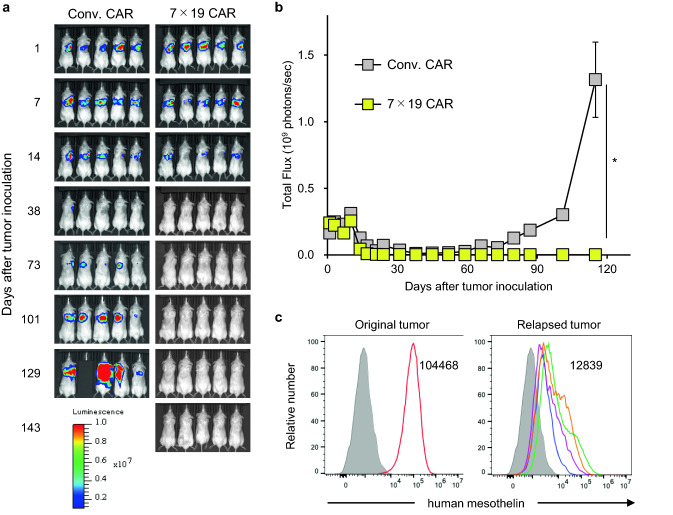
Fig. 4.
Prevention of orthotopic tumor relapse by the treatment with 7 × 19 CAR-T cells Immunodeficient NSG mice were intrapleurally inoculated with 2 × 10⁶ ACC-MESO1-GFP-Luc cells on day 0, followed by i.v. injection of 1 × 10⁵ Conv. CAR-T or 7 × 19 CAR-T cells on day 1. Tumor growth was assessed using IVIS every week. a Representative bioluminescence images of the mice are shown. b Total flux of whole-body bioluminescence measured by IVIS is shown as mean ± SEM (representative data from two independent experiments). c Relapsed ACC-MESO1-GFP-Luc tumor cells were harvested from the chest cavity of the mice, which were treated with Conv. CAR-T cells and euthanized on day 79–126 due to massive tumor relapse, and individually assessed for the expression of endogenous mesothelin using flow cytometry (right panel, open histograms with four distinct colors indicating four individual relapsed tumors). As a control, the expression level of endogenous mesothelin in the original ACC-MESO1-GFP-Luc tumor cells was also examined (left panel, open histogram). The filled histogram indicates an unstained control. The numbers in the histogram indicate the mean fluorescence intensity of mesothelin expression in the original and relapsed tumors (average of four individual relapsed tumors in right panel)

