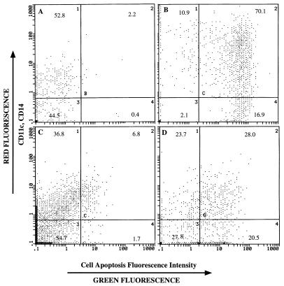
FIG. 7.
Apoptosis of macrophages pulsed with viral polypeptides induced by CD4+ CTL. All macrophages were treated with anti-macrophage antibodies (anti-CD11c and anti-CD14) and were gated by forward and low-angle light scatter as large and complex cells. The anti-CD11c and anti-CD14 antibodies were labeled with phycoerythrin (red fluorescence) to identify macrophages, while the fragmented DNA of apoptotic cells were labeled with fluorescein isothiocyanate-dUTP by terminal deoxynucleotidyltransferase (green fluorescence). Intact macrophages were either not labeled with fluorescein isothiocyanate-dUTP (negative control) (A) or not labeled with fluorescein isothiocyanate-dUTP (positive control) (B). Compared to the target cells without viral polypeptide treatment (medium control) (C), about 30% of target cells pulsed with BHV-1 polypeptides exhibited apoptosis in CTL assays (D).

