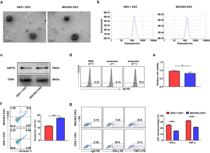
Fig. 3.
GC cell-derived exosomes regulated the function of Vγ9Vδ2 T cells. a–c GES-1 cell-derived exosomes (GES-1 EXO) and MGC803 cell-derived exosomes (MGC803 EXO) were identified by TEM a, NTA b and western blotting c. The expression of exosomal markers HSP70 and CD63 was analyzed by western blotting. (d) The proportions of Dil-positive Vγ9Vδ2 T cells among Vγ9Vδ2 T cells treated with Dil-labeled exosomes at 4 ℃ or 37 ℃ were detected by flow cytometry. e The cell viability of Vγ9Vδ2 T cells treated with exosomes from GES-1 (GES-1 EXO) or MGC803 (MGC803 EXO) cells was analyzed by CCK-8 assay. f, g The apoptosis rate (f) and the IFN-γ and TNF-α production g of Vγ9Vδ2 T cells treated with exosomes from GES-1 (GES-1 EXO) orMGC803 (MGC803 EXO) cells were detected by flow cytometry. Values are expressed as means ± SD. Data are representative of results from three independent experiments. *p < 0.05, **p < 0.01, ***p < 0.001

