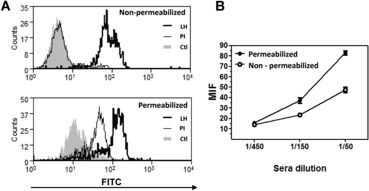
Fig. 3.
Antibodies induced by HCF immunization recognize tumor cells. a Histogram plots showing antibody recognition for membrane and cytosolic antigens on LL/2 cells. Flow cytometry analyses were carried out on permeabilized or non-permeabilized LL/2 tumor cells incubated with sera (diluted 1:100) collected from animals immunized with human HCF. Controls consisted on pre-immune sera (PI) or cells incubated with secondary antibody only (Ctl). Five thousand events were collected and gated on FSC vs SSC dot plot. b Mean fluorescence intensity (MFI) representing antibody recognition of membrane and cytosolic antigens on non-permeabilized and permeabilized LL/2 cells, respectively. In this case, different sera dilutions (1:50, 1:150, 1:450) were used, and MFI values were subtracted to the corresponding pre-immune serum at same dilution

