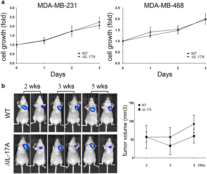
Fig. 1.
The role of IL-17A on cell growth in MDA-MB-231 and MDA-MB-468 cell lines. After IL-17A was knocked down in MDA-MB-231 and MDA-MB-468 cells (a), cell proliferation was evaluated by trypan blue exclusion assay. Wild type (WT) and knocked down IL-17A (∆IL-17A) MDA-MB-231 cells (1 × 10⁷ in 0.1 mL PBS) containing a luciferase gene were injected into back of immunodeficient NU-Foxn1nu mice. At the 2nd, 3rd, and 5th week, the progression of the tumors was visualized using an in vivo imaging system (IVIS) and tumor volume was quantified (b). Two-way ANOVA was used for statistical analysis for cell growth in vitro and in vivo

