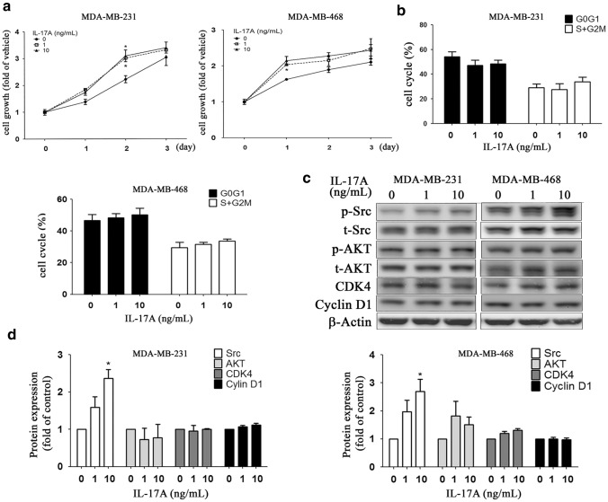
Fig. 2.
Effects of exogenously administration of IL-17A on cell growth in MDA-MB-231 and MDA-MB-468 cell lines. MDA-MB-468/231 cells were cultured in low serum medium with a cell density (1 × 10⁴ / well), followed by treatment of different doses of IL17A (0-, 1-, 10 ng/mL). After 1, 2, and 3 days of treatment, cell proliferation rate was evaluated by trypan blue assay (a). For cell cycle analysis, cells (2 × 10⁵ / well) were cultured for 24 h in low serum medium, followed by another 24 h-culture, and then cells were harvested for cell cycle analysis (b). Cell cycles were presented as percentages of cell cycle fraction, namely sub-G0/G1 phase, G0/G1 phase, S phase, and G2/M phase. Growth-related signaling proteins expression, such as Src, AKT, CDK4, and cyclinD1, were analyzed by Western blot (c) and quantified (d). Asterisk indicates a p value < 0.05 (one-way ANOVA)

