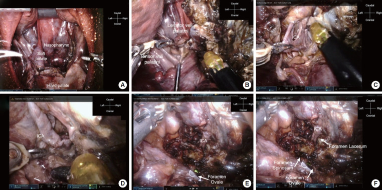
Fig. 3.
da Vinci Xi Surgical System (Intuitive Surgical, Inc., Sunnyvale, CA, USA) approach to the skull base in the coronal plane. (A) View after lateralization of the soft palate. (B) Dissection of the tensor veli palatini and levator veli palatini muscles. (C) Identification of the ICA to avoid injury to the soft connective tissue. (D) Following the ICA to the skull base. (E) Dissection of the soft connective tissue. The ICA and foramen ovale can be identified; the white tube shows the foramen ovale. (F) The ICA going through the foramen lacerum into the intracranial space. The reachable lateral limitation is the foramen ovale. ICA, internal carotid artery.

