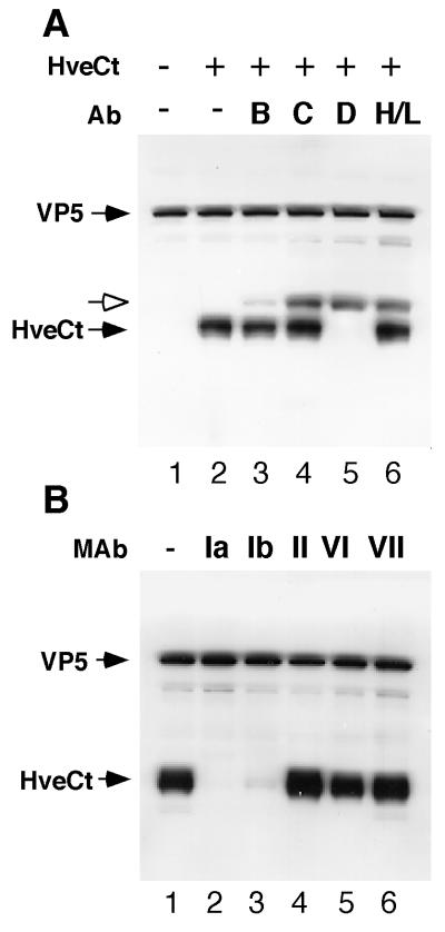
FIG. 8.
Binding of HveCt to HSV particles is blocked by anti-gD antibodies. (A) Purified HSV-1 KOS virions (107 PFU) were incubated at 4°C for 2 h with (lane 2) or without (lane 1) HveCt (150 μg) and loaded onto a sucrose gradient. The viral band was collected and analyzed by SDS-PAGE and Western blotting. Membranes were probed for presence of VP5 and HveCt (R154 serum). In blocking experiments, virions were preincubated with cocktails of antibodies (Ab) specific for HSV glycoproteins: for gB, SS10 (0.5 μl of ascites), DL16 (5 μg of IgG), DL21 (5 μg of IgG), and R69 (0.5 μl of serum) (lane 3); for gC, MP1 (0.5 μl of ascites), MP5 (5 μg of IgG), 1C8 (5 μg of IgG), and R46 (0.5 μl of serum) (lane 4); for gD, 1D3 (0.5 μl of ascites), DL2 (5 μg of IgG), DL11 (5 μg of IgG), and R7 (0.5 μl of serum) (lane 5); for gH-gL, LP11 (0.5 μl of ascites), 53S (5 μg of IgG), H6 (5 μg of IgG), and R137 (0.5 μl of serum) (lane 6). Rabbit Ig heavy chain is detected by goat anti-rabbit secondary antibody and is indicated with a white arrow. (B) Prior to cosedimentation with HveCt, purified HSV-1 KOS virions (107 PFU) were preincubated with 50 μg of a monoclonal IgG (HD1 [group Ia], DL11 [group Ib], DL6 [group II], DL2 [group VI], or 1D3 [group VII]) during 1 h at 37°C. Untreated control is shown in lane 1.

