Fig. 2.
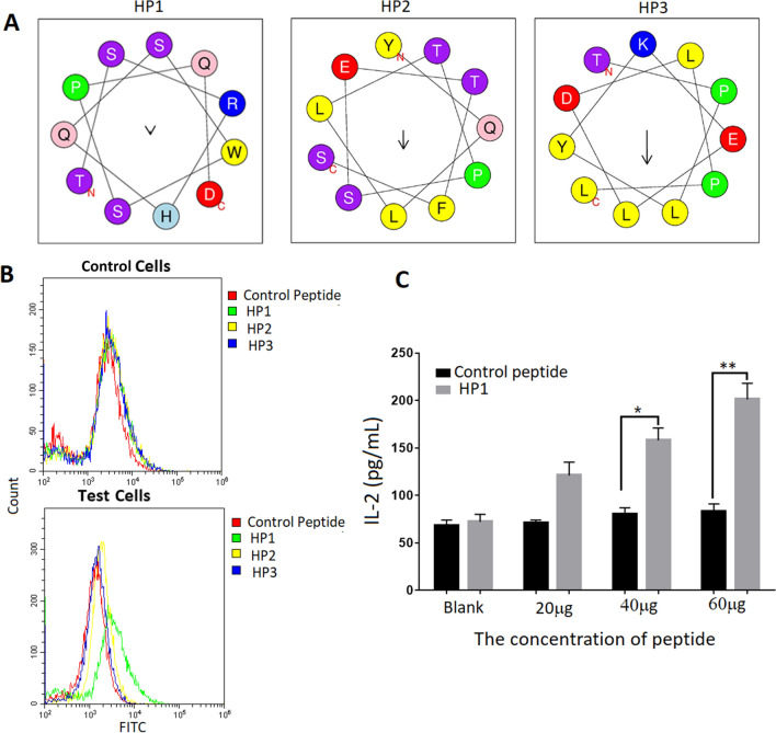
Confirmation of the peptide’s capability to bind to HCC-specific transfected cells. a The spiral structure of three peptides were predicted by the Heliquest website http://heliquest.ipmc.cnrs.fr/?tdsourcetag=s_pcqq_aiomsg. Enter the website, click the button of analysis, enter the sequence of HP1, HP2 and HP3 in the blank. Helix type is 3–10, and click the button of process. The predicted spiral structure of the three peptides was presented. b HP1 was able to bind to test cells by FACS analysis. Three peptide and control peptide conjugated with FITC (10 μg) were co-cultured with test cells and control cells, respectively. The results showed that HP1 was better able to bind to test cells than that of the control cells (Green line represents HP1). Results are representative of three independent experiments. c HP1 could also stimulate test cells to secrete IL-2 in a dose-dependent manner. Different dosages of HP1 and control peptide were co-cultured with test cells and control cells for 24 h. Production of IL-2 in the supernatant of cell culture medium was detected by ELISA. Data are shown as the mean of three independent experiments. *Denote P < 0.05. **Denote P < 0.01

