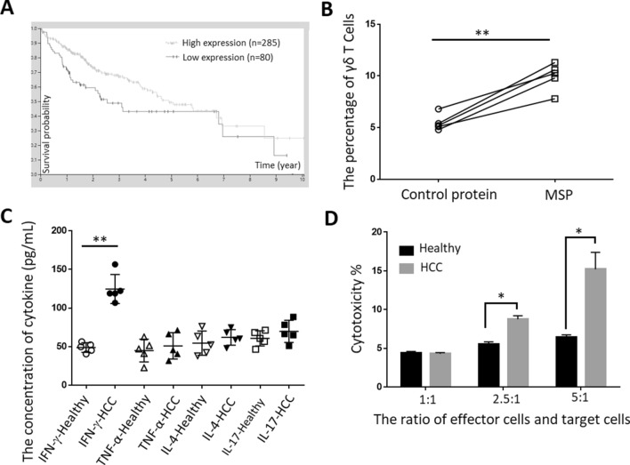
Fig. 4.
Effector function of MSP-induced γδT cells from HCC patients. a The relationship of MSP with the survival probability of HCC patients, as shown by the OncoLnc website. In the oncolnc website, MSP was entered to get the Cox regression results. HCC is selected, lower percentile was set on 10 and higher percentile was set on 90, the Kaplan plot for MSP in HCC was determined. b Immobilized MSP could induce proliferation of γδT cells from HCC patients. Immobilized MSP and control protein were pre-coated onto microtiter plates, on which γδT cells from 5 HCC patients were incubated for 2 weeks. The percentage of γδT cells was measured by FACS. c MSP-activated T cells could secrete Th1-based cytokines. Immobilized MSP was pre-coated on microtiter plates, on which γδT cells from 5 HCC patients and 5 healthy controls were added. Supernatants were collected after 48 h and measured for IFN-γ, TNF-α, IL-4 and IL-17 secretion. d MSP-induced γδT cells from HCC patients have the cytotoxicity of to HepG2 by CytoTox 96 Non-radioactive Cytotoxicity Assay. After γδT cells from HCC patients inoculated by MSP were sorted and cultured, cytotoxic activities of γδT cells against HepG2 in vitro was evaluated. Data are shown as the mean of three independent experiments. *Denote P < 0.05. **Denote P < 0.01

