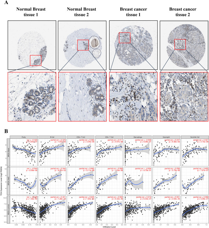
Fig. 5.
TOX localization and relation to tumor infiltrating immune cells in breast cancer. a Representative immunohistochemical staining of TOX protein in two normal breast tissues and two breast cancer tissues. Images were extracted with quantification from Human Protein Atlas. Pathological observations and quantification data for all analyzed tissue images for normal breast and tumor tissues have been given in Supplementary Tables S1 and S2, respectively. b Correlation of TOX gene expression with tumor purity and six major tumor infiltrating immune cell types in different breast cancer subtypes from TCGA-BRCA dataset

