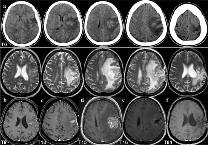
Fig. 1.
Patient brain neuroimaging. a July 2013 (Time 0): pre-surgery brain computed tomography (CT) showing an intra-axial frontal expansive lesion with edema and mass effect. b–f Brain MRI, top T2 and bottom T1 with contrast agent weighted imaging, respectively; b April 2014 (Time 9): MRI after gross total resection (GTR) of the lesion, slight gliosis without mass effect; c, d August–October 2014 (Time 13–Time 15): recurrence with T1 enhancing lesion with increasing size, edema and mass effect; e November 2014 (Time 16): early brain MRI post-second surgery and GTR of the rGBM, showing persisting edema and mass effect; f June 2020 (Time 84): brain MRI shows gliosis and ex-vacuum right lateral ventricle enlargement, with no signs of recurrence

