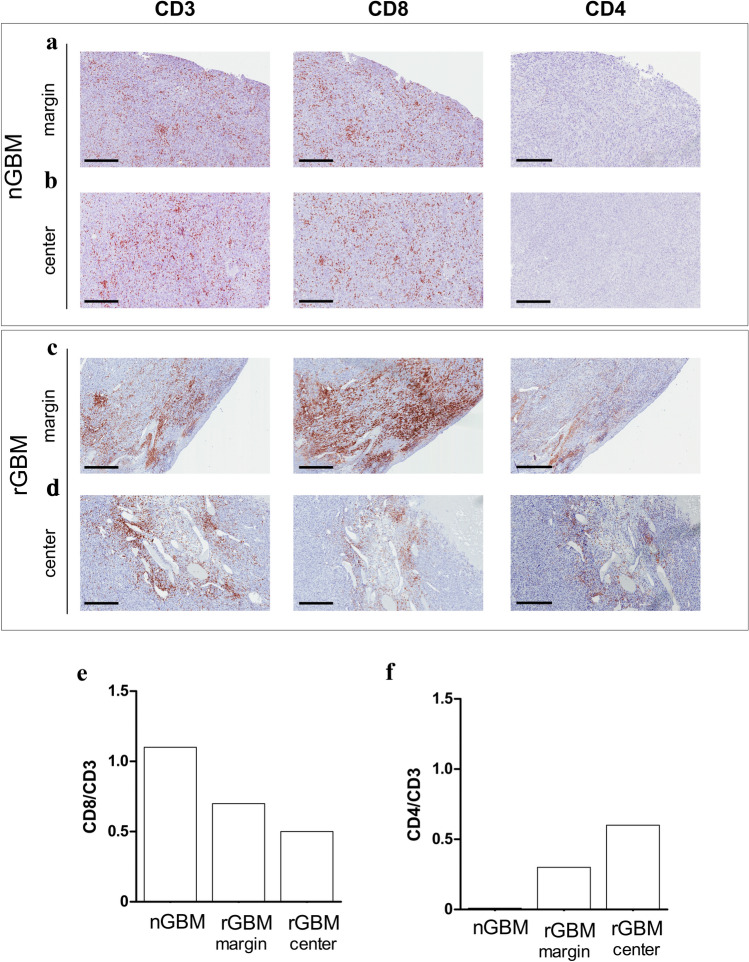
Fig. 3.
CD8 + cell infiltration are abundant in both nGBM and rGBM specimens. a, b Representative adjacent sections of the nGBM specimen showing a high distribution of CD3 + and CD8 + and total absence of CD4 + TILs. High density of CD3 + and CD8 + TILs was observed in both tumor center (a) and tumor margins (b) of the nGBM specimen. c, d Changes in CD3 + and CD8 + T cell distribution and increased of CD4 + TILs were found in the rGBM. A manual count performed in the center area and tumor margins separately confirmed a significant higher density of CD3 + CD8 + TILs at the tumor margins (Scale Bar 300 μm)

