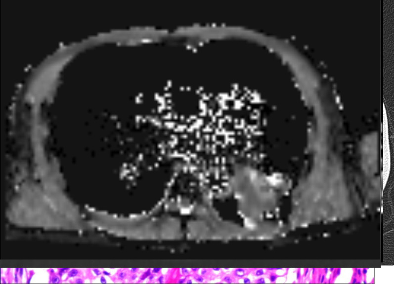
Figure 4.
A representative case of pulmonary masses. A 58-year-old male confirmed as squamous cell carcinoma in the left lower lung zone by surgery. (a) CT image. (b) DWI image (b = 800 s/mm2). (c) ADC map. (d) IVIM-D map. (e) IVIM-D* map. (f) IVIM-f map. (g) The generated curve of IVIM. (h) Photomicrograph of the lesion (HE × 200). The measured D value was 1.21 × 10⁻³ mm2/s, indicating a malignant tumour, whereas the measured ADCmean (1.56 × 10⁻³ mm2/s) was obviously higher, suggesting that it was influenced by the perfusion effect. ADC, apparent diffusion coefficient; DWI, diffusion-weighted imaging; IVIM, intravoxel incoherent motion.

