Fig. 2.
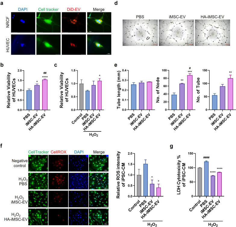
Multifunctional effects of HA-iMSC-EVs on the various cell types. a Incorporation of DiD-labeled HA-iMSC-EVs into NRCF and HUVEC. The uptake of DiD-labeled HA-iMSC-EVs was examined after 24 h treatment with 100 μg/mL EVs (600 × magnification). b–c Enhanced effect of HA-iMSC-EVs on endothelial cell proliferation. Comparison of the viability of HUVEC (b) and H2O2-damaged HUVEC (c) after treatment with PBS, iMSC-EVs, or HA-iMSC-EVs. Data are presented as mean ± SD (n = 6 and n = 3, respectively). *p < 0.05; ****p < 0.0001 vs PBS; ## p < 0.01 vs iMSC-EV; one-way ANOVA. d–e Tube formation by HUVEC treated with iMSC-EVs or HA-iMSC-EVs. d Representative images of HUVEC tube formation. e Quantification of tube length (left), number of nodes (middle), and number of tubes (right). Data are presented as the mean ± SD (n = 3). **p < 0.01; ***p < 0.001 vs PBS; # p < 0.05 vs iMSC-EV; ns, not significant; one-way ANOVA. f–g Cytotoxicity reducing effects of HA-iMSC-EVs on hiPSC-CMs. f Representative image and fluorescence intensity quantification graph of ROS levels in hiPSC-CMs with H2O2-induced oxidative damage after treatment with PBS, iMSC-EVs, or HA-iMSC-EVs. *p < 0.05 vs PBS. g Comparison of LDH levels representing cytotoxicity in H2O2-injured hiPSC-CMs after treatment with PBS, iMSC-EVs, or HA-iMSC-EVs. Data are presented as the mean ± SD (n = 3). ****p < 0.0001 vs PBS; #### p < 0.0001 vs Control; one-way ANOVA

