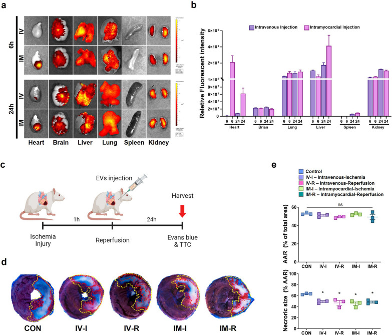
Fig. 6.
Cardiac protection effect of HA-iMSC-EVs against myocardial ischemia–reperfusion injury. a, b In vivo tracking of HA-iMSC-EVs. The localization of fluorescently labeled HA-iMSC-EVs was visualized after 6 or 24 h of intravenous (IV, upper) and intramyocardial (IM, lower) administration (a). b Bar graph shows the relative fluorescent intensity. c Experimental Process. HA-iMSC-EVs (20 mg/kg) were injected intravenously or intramyocardially 5 min before or after reperfusion. d Representative images of TTC/Evan’s blue staining 24 h after reperfusion. e The area at risk (AAR) was not significantly different between the I/R control and HA-iMSC-EVs injection groups, but necrotic size (white) was significantly reduced in the HA-iMSC-EVs injection group. * p < 0.05, compared to the PBS treatment group by unpaired t-test. Error bars show means ± SEM. n = three animals per group. ns, not significant

