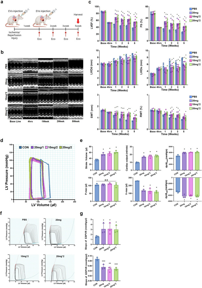
Fig. 7.
Injection of HA-iMSC-EVs improved cardiac function and reduced adverse cardiac remodeling. a Experimental Process. HA-iMSC-EVs (10 mg/kg or 20 mg/kg) were injected intramyocardially 5 min before reperfusion. The second injection was administered 1 week after the first treatment. b Representative M-mode echocardiography images at baseline (4 h) and 1, 2, and 5 weeks after EVs injection. c Differences in left ventricular ejection fraction (EF), fractional shortening (FS), septal wall thickness (SWT), end-diastolic (LVIDd), and end-systolic dimensions (LVIDs) between baseline and 5 weeks after EVs injection. * p < 0.05, compared with the PBS treatment group by one-way ANOVA. Error bars show means ± SEM. n = 4–5 animals per group. d Representative pressure–volume loops obtained from catheter-based left ventricular P–V measurements five weeks after IR injury. e Cardiac output, stroke volume, volume max, pressure max, and systolic and diastolic functions as measured by the maximal and minimal rates of pressure change during isovolumic relaxation (dP/dtmax and dP/dtmin). f Measurement of cardiac contractibility through inferior vena cava occlusion. g Slope of end-systolic pressure–volume relationship (ESPVR) and end-diastolic pressure–volume relationship (EDPVR). * p < 0.05; **p < 0.01; ***p < 0.001, compared with the PBS treatment group by Bartlett test in R. Error bars show means ± SEM. n = three animals per group

