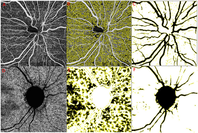
Figure 1.
Example images for retinal layer segmentation and large vessel removal. RPC layer blood flow diagram (A–C), original optic disc margin threshold (A), optic perfusion areas (B), and the major branch retinal blood vessels (C). Choroidal capillary blood flow diagram (D–F), original chorioidal capillary layer (A), no-perfusion areas (E), and the major branch retinal blood vessels and optic disc (F). RPC, radial peripapillary capillaries.

