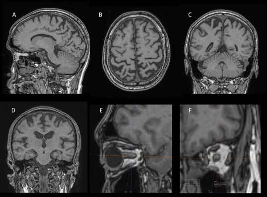
Figure 1.

Selected T1-MPRAGE images demonstrating sagittal (A), axial (B), and coronal (C) images used for PCA scoring, selected coronal image (D) for MTA scoring, and focused images (E & F) for optic nerve surface area measurement. The above images show PCA scoring of 2 and MTA score of 0. MPRAGE, magnetization-prepared rapid gradient-echo; MTA, medial temporal atrophy; PCA, posterior cortical atrophy.
