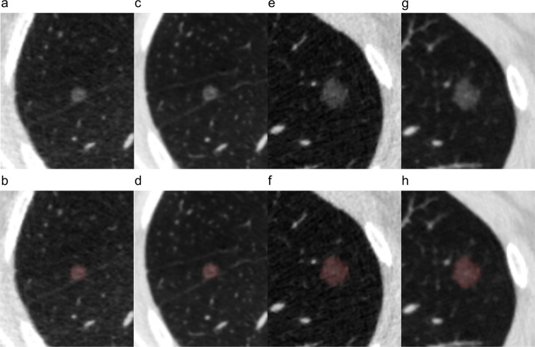
Figure 2.
Representative images and segmentation results of nodules. (a, b, c, d) A 53-year-old male with minimally invasive adenocarcinoma. (e, f, g, h) A 41-year-old female with invasive adenocarcinoma. (a, e) Original images and (b, f) segmentation results of low-dose CT. (c, g) Original images and (d, h) segmentation results of standard-dose CT.

