Figure 3.
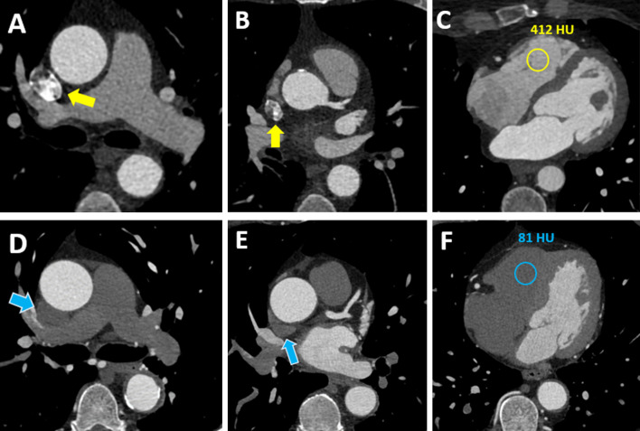
66 year-old female with chest pain underwent standard CCTA (Case 1, (A-C) and 65-year-old male with known coronary artery disease and recurrence of angina underwent CCTA using the test bolus evaluation algorithm (Case 2, (D-F). Axial CCTA sections are shown. Beam hardening artifact is shown in the superior vena cava in case one with standard CCTA (A, B; yellow arrows), while artifact is not present in Case two with test bolus algorithm based CCTA (D, E; blue arrows). Furthermore, panel (C) shows a contrast filled right ventricle with a density of 412 HU. In contrast, panel (F) demonstrates low attenuation in the right ventricle (81 HU). CCTA, coronary computed tomography angiography; HU = Hounsfield Units.

